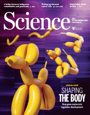
배아줄기세포가 분화하면서 우리 몸이 형성될 때, 유전자의 발현량에 따라 발달 과정의 모습을 풍선아트에 비유한 사이언스 표지 그림에 관한 내용입니다.
(원문)

Science 제공
풍선을 불어 개나 고양이, 꽃 모양 등을 만드는 것을 ‘풍선 아트’라 부른다. 그런데 부는 세기가 약간만 어긋나도 다리나 허리 부분이 비정상적으로 긴 개가 만들어지곤 한다.
국제학술지 ‘사이언스’는 29일 점점 개모양을 갖춰가는 풍선의 모습을 표지로 소개했다. 배아줄기세포가 분화하면서 우리 몸이 형성될 때, 유전자의 발현량에 따라 발달 과정의 모습을 풍선아트에 비유한 것이다.
이번 호에서는 유전자 발현과 관련해 쥬느비에브 알모츠니 프랑스 파리 퀴리연구소 책임연구원 연구진은 ‘염색체 가소성: 세포 운명과 정체성을 결정하는 풍경’이라는 제목의 논문과 종유안 루오 미국 솔크연구소 교수 연구진이쓴 ‘역동적인 DNA 메틸화: 필요한 시간과 정확한 위치에서’라는 리뷰 논문 등 논문 다섯 편이 특집으로 공개됐다.
생명체의 밑그림에 해당하는 유전자가 발현해 몸을 구성하려면 크게 두 가지가 제대로 작동해야 한다. 하나는 염색체가 붙어 있는 히스톤 단백질에 메틸기가 제때에 붙어 유전자가 작동할 수 있도록 풀리는 것이다. 다른 하나는 풀린 염색체 위에 적절한 전사인자가 붙어 DNA 염기가 해독되면서 단백질을 형성해야 한다.
알모츠니 책임연구원은 “다분화 능력이 있는 배아 세포가 특정 조직으로 성장하려면 정체성을 갖춰야 한다”며 “히스톤 단백질의 메틸화나 전사인자 등 다양한 변수를 연구해야 한다”고 밝혔다. 그는 “최근 생체 내부를 관찰하는 기술이 발전하면서 발달 과정에서 일어나는 염색체의 변화에 대한 심층적인 연구가 진행되고 있다”고 덧붙였다.
루오 교수는 자신의 논문에서 “단일 배아세포를 측정하는 기술과 이들에 관여하는 유전자를 골라내는 기술 진보가 이뤄지고 있다”며 “특정 메틸기가 염색체에 작용하는 현상을 보다 확실히 연구하게 됐다”고 말했다.
Hox genes and body segmentation
Science 28 Sep 2018:
Vol. 361, Issue 6409, pp. 1310-1311
DOI: 10.1126/science.aav0692
원문: 여기를 클릭하세요~
On page 1377 of this issue, He et al. (1) elucidate two long-standing problems in animal evolution: the ancient function of the homeobox (Hox) gene cluster, which has puzzled scientists for decades (2), and the centuries-old debate on the emergence of the segmented animal body.
Hox genes were first discovered in flies and mice, where they specify different body segments along the anterior-posterior (A-P) axis (3). Although their expression often overlaps in posterior body regions, they show spatially distinct anterior expression boundaries (4) (see the figure). Importantly, the A-P sequence of Hoxgene expression in the body matches their 3′ to 5′ sequential occurrence within a chromosome cluster, a principle called spatial collinearity. Moreover, the more anteriorly expressed 3′ Hox genes are often expressed earlier in development, which is called temporal collinearity. In addition, individual Hox proteins are typically active close to their anterior expression boundary, because the more 5′, or posterior, proteins counteract the function of the more 39, or anterior, ones whenever both products co-exist. This is called posterior prevalence (4).
Making sense of these rules has been challenging. For vertebrates, Hox collinearity and posterior prevalence are explained with the sequential activation of these genes from within a posterior growth zone (5). This growth zone iteratively produces mesodermal body segments, called somites, and pushes them toward the anterior. If, during this process, Hox genes are sequentially activated and stay on in the newly generated segments, then staggered expression of Hox genes arises along the A-P axis.
What is the relevance of spatial collinearity for invertebrates? Collinear Hox gene activity is described for patterning ectodermal derivatives such as the nervous system in insects (3). Few studies revealed Hox gene expression in mesodermal structures that resemble vertebrate somites. Moreover, Hox gene clusters are active in both segmented and unsegmented invertebrates such as sea urchins (6). This has prompted the view that the tight link between Hox genes and body segmentation observed in vertebrates, insects, or annelids has evolved independently, that is, by evolutionary convergence (7).
Hox genes in sea anemone and mouse development
Collinear expression of gastrulation brain homeobox (Gbx), “anterior” Hox (Anthox6a), “middle” Hox (Anthox8), and “posterior” Hox(Anthox1a) genes defines gastric pouches in sea anemone, and that of HoxA genes defines somite positioning in mice.
GRAPHIC: ADAPTED BY V. ALTOUNIAN/SCIENCE FROM D. ARENDT
The best way to challenge this view is to investigate an evolutionary outgroup. Accordingly, He et al. investigated Hox gene function in the cnidarian Nematostella vectensis, the starlet sea anemone. Cnidarians are our most distant relatives to possess a Hox cluster (see the figure). Their inner surface is folded, so that their primitive gut is subdivided into chambers, called gastric pouches. These are also continuous with the lumen of the tentacles. The cnidarian lineage diverged from ours when a cluster of only three Hox genes existed (8), with one anterior (3′), one middle, and one posterior (5′) gene. The sea anemone has a fragmented version of the ancient three-gene Hox cluster, with additional, lineage-specific duplications (1). Previous expression comparisons revealed expression of N. vectensis Hox genes in the developing gastric pouches, with distinct Hox gene expression boundaries matching the epithelial boundaries between the pouches (9–11). These boundaries are collinear with the assumed sequence of genes in the cnidarian-bilaterian ancestor [referred to as trans-spatial collinearity (4)].
He et al. established gene ablation techniques in N. vectensis to investigate the function of the Hox genes. They uncovered a fundamental role of these gene products in the formation of the gastric pouches. In the absence of Hox activity, the epithelial folds separating these pouches are lost. Therefore, similar to vertebrates, N. vectensis Hox genes differentially and specifically control the generation of boundaries between repeated body parts, in conjunction with a later role in controlling the different fates of these parts (12). He et al. also show that the N. vectensis Hox genes are expressed sequentially, in concert with the stepwise appearance of folds during polyp development. Thus, N. vectensis Hox genes show spatial and temporal collinearity. Additionally, the loss of each Hox gene abolishes the epithelial folding at the most anterior expression limit of that gene—where they are not coexpressed with a more posterior Hox gene. In the absence of the boundary formed by the folds, the two adjacent pouches fuse. These phenotypes are consistent with the posterior prevalence principle.
This is strong evidence that the link between Hox gene function and some kind of body segmentation is ancestral. But how does N. vectensis segmentation—the sequential generation of gastric pouches by epithelial folding—relate to bilaterian segmentation? The simple mode of epithelial folding observed in cnidarians has inspired morphologists for more than 150 years, who saw similarities with mesodermal segmentation in bilaterians. Bilaterian mesodermal somites often emerge as outpocketings from the gut. The enterocoel theory proposed that both cnidarian pouches and bilaterian somites are derived from such outpocketings (7, 13). The findings of He et al. support this theory.
One possible caveat is that the sequentially emerging N. vectensis folds are not generated from a posterior growth zone. This might represent a secondary simplification of cnidarian development, given that in the tube anemones (Cerianthida), which represent an early diverging branch of Cnidaria, additional folds are pushed forwards from a terminal growth zone (9). Examining the expression of Hox genes and of growth zone markers in these cnidarians could be especially rewarding, as it might establish a similar link between Hox spatial and temporal collinearity and the generation of body segments from a growth zone as is observed in vertebrates (4, 5). Another note of caution concerns the unsolved axial relationships between cnidarians and bilaterians (14), which led to conflicting views about the nature of the cnidarian Hox axis (15). However, the data of He et al. seem to firmly settle this issue.
Twenty-five years after the revolutionary zootype concept, which considered the Hox genes a shared feature of all animals (2), we can now refine this idea and propose that the Hox gene cluster evolved with the emergence of segmental epithelial folds and pouches in the cnidarian-bilaterian ancestor. These persisted as gastric pouches in today’s cnidarians and gave rise to mesodermal somites in the bilaterians.
