Abstract
Recent studies have demonstrated an array of stem cell–derived, self-organizing miniature organs, termed organoids, that replicate the key structural and functional characteristics of their in vivo counterparts. As organoid technology opens up new frontiers of research in biomedicine, there is an emerging need for innovative engineering approaches for the production, control, and analysis of organoids and their microenvironment. In this Review, we explore organ-on-a-chip technology as a platform to fulfill this need and examine how this technology may be leveraged to address major technical challenges in organoid research. We also discuss emerging opportunities and future obstacles for the development and application of organoid-on-a-chip technology.
Decades of study in developmental biology and stem cell research have advanced our ability to recapitulate the key aspects of organogenesis in vitro. Recent years have seen considerable progress toward exploiting the self-organizing properties of pluripotent or adult stem cells to generate organotypic multicellular constructs known as organoids (1, 2). Thanks to their ability to emulate microarchitecture and functional characteristics of native organs, organoids are emerging as a promising approach for the modeling of development, homeostasis, and disease of various human organs (1–3).
The conventional methods of forming organoids rely on three-dimensional (3D) culture of mammalian stem cells with sequential addition of growth factors. Although this approach has been widely used because of its simplicity, there is growing recognition that the current organoid culture techniques have the potential for substantial improvement. In particular, the random configuration of traditional 3D culture makes it difficult to precisely control organoids and their local environment. Existing culture systems also have limited capacity to reproduce the complex and dynamic microenvironment of a developing organ that provides instructive cues for organogenesis (4, 5). The lack of these environmental signals poses challenges to achieving more complete, in vivo–like organoid development in a reproducible manner (3, 6).
To address the limitations of conventional culture techniques, researchers in stem cell and developmental biology are forming alliances with engineers and physical scientists to develop advanced in vitro technologies for organoid research. At the forefront of this undertaking is the integration of organoids with organ-on-a-chip technology.
What are organs-on-a-chip?
Organs-on-a-chip can be broadly defined as microfabricated cell culture devices designed to model the functional units of human organs in vitro (7–12). In general, the construction of any organ-on-a-chip system is guided by design principles based on a reductionist analysis of its target organ (Fig. 1). The first step is to understand the anatomy of the target organ and reduce it to the basic elements essential for physiological function. These functional units are then examined to identify key features such as different cell types, structural organization, and organ-specific biochemical and physical microenvironments. For example, the alveolar–capillary unit of the lung consists of alveolar epithelial cells (cell type 1) and pulmonary microvascular endothelial cells (cell type 2) that are closely apposed to each other and separated by a thin interstitium (structural organization) (Fig. 1A). The epithelial and endothelial layers are subjected to air and blood flow, respectively, and the multilayered interface experiences breathing-induced cyclic mechanical stretch (organ-specific microenvironment).
(A) Reductionist analysis of a target organ (lung) identifies alveoli as the functional unit composed of epithelial and endothelial cells separated by a thin interstitium. (B) An analogous model is constructed from three layers to bring these two cell types into physiological proximity. (C) To mimic breathing-induced mechanical activity, the cells are cyclically stretched by applying vacuum (vac) to the side chambers. [Illustration: BIOLines Lab]
Next, a cell culture device is designed to replicate the identified features. The device often contains multiple, individually addressable flow-through microchambers to grow multiple cell types while controlling the culture environment in a cell type–dependent manner. If necessary, additional components are incorporated that can be actuated mechanically, chemically, electromagnetically, or optically to emulate the biochemical and mechanical environment of the target organ. Finally, the designed device is produced using microfabrication techniques such as soft lithography (13).
The design strategy outlined here has been successfully implemented to create an organ-on-a-chip model of the alveolar–capillary unit of the lung (14). This system consists of two overlapping microchannels separated by a thin, flexible, microporous membrane (Fig. 1B, left). The compartmentalized design enables coculture of alveolar epithelial cells and lung microvascular endothelial cells on either side of the membrane while the cells are exposed to their respective tissue-specific environment (i.e., air on the alveolar side and fluid flow on the vascular side) (Fig. 1B, right). To mimic the deformation of the alveolar–capillary interface during breathing, the device is also equipped with two hollow microchambers alongside the culture channels, in which cyclic vacuum application induces stretching of the cell-lined intervening membrane (Fig. 1C).
By integrating living human cells with synthetically generated yet physiologically relevant microenvironments, organs-on-a-chip can mimic integrated organ-level functions necessary for physiological homeostasis, as well as complex disease processes (15, 16). Furthermore, different organ-chip models can be fluidically linked to construct “body-on-a-chip” systems capable of simulating multiorgan interactions and physiological responses at the systemic level (17, 18). Although these advanced model systems are still far from achieving the functionality of real human organs, their ability to capture key aspects of human physiology and pathophysiology makes them a promising approach for complementing and reducing animal studies for preclinical assessment of drugs, medical devices, and biomaterials (7, 9, 19). Organ-on-a-chip technology also provides an attractive in vitro platform for screening adverse health effects of chemicals, environmental materials, and consumer products (10, 20).
(원문: 여기를 클릭하세요~)
Cancer modeling meets human organoid technology
Abstract
Organoids are microscopic self-organizing, three-dimensional structures that are grown from stem cells in vitro. They recapitulate many structural and functional aspects of their in vivo counterpart organs. This versatile technology has led to the development of many novel human cancer models. It is now possible to create indefinitely expanding organoids starting from tumor tissue of individuals suffering from a range of carcinomas. Alternatively, CRISPR-based gene modification allows the engineering of organoid models of cancer through the introduction of any combination of cancer gene alterations to normal organoids. When combined with immune cells and fibroblasts, tumor organoids become models for the cancer microenvironment enabling immune-oncology applications. Emerging evidence indicates that organoids can be used to accurately predict drug responses in a personalized treatment setting. Here, we review the current state and future prospects of the rapidly evolving tumor organoid field.
(원문: 여기를 클릭하세요~)
항암·맞춤치료 혁명 가져올 ‘오가노이드’

실제 조직이나 장기와 닮은 오가노이드를 이용하면 병의 원인이나 진행 과정을 밝히거나 신약의 효과 등을 확인할 수 있다. 사이언스 제공
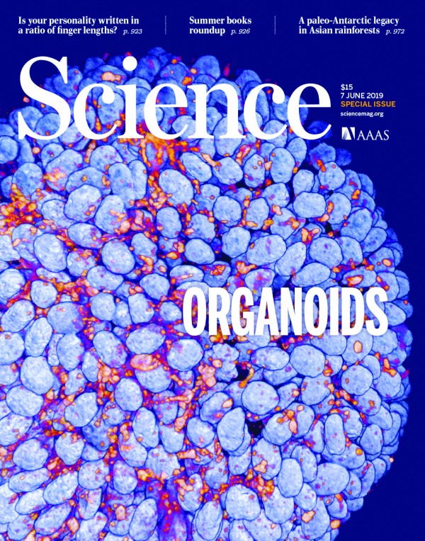
국제학술지 사이언스는 7일 인간의 기도를 흉내 낸 오가노이드를 3차원 공초점현미경으로 촬영한 사진을 표지로 담았다. 파란색이 DNA, 빨간색이 단백질이다. 사이언스는 이번 특집기사에서 십수년간 과학자들이 오가노이드를 만들어 연구한 결과를 총정리했다.
오가노이드는 줄기세포를 3차원으로 쌓아 배양한 것으로 실제 조직과 닮아 약물 효과나 질병 원인, 생리적인 반응 등을 연구할 때 사용한다. 동물에게 실험할 때보다 훨씬 정확한 결과를 얻을 수 있고, 살아 있는 사람에게 할 수 없는 실험까지 할 수 있다는 장점이 있다. 오가노이드가 인체를 대체하기 위해서는 실제 세포 내에서 일어나는 현상과 미세환경이 닮아야 하고, 외부에서 인위적으로 조절할 수 있어야 한다.
먼저 한스 클레버스 네덜란드 위트레흐트 의학연구소 교수와 데이비드 투베슨 미국 로스가르텐 연구재단 췌장암 연구소 수석 연구원 공동연구팀은 그간 과학자들이 암을 연구하기 위해 오가노이드를 만들었던 연구 결과를 소개했다. 암이 발생하는 원인을 찾거나, 암이 전이되는 과정, 또는 새로 개발한 항암제의 효과를 확인할 때 오가노이드를 쓸 수 있다.
클레버스 교수는 “체내에서 암이 발견되면 빨리 없애야 하는데, 암에 대해 연구하려면 오랫동안 살아 있는 암 조직 샘플이 많이 필요하다”면서 “암조직을 흉내 낸 오가노이드가 확실한 도구”라고 말했다. 그는 “예를 들어 MLH1, APC처럼 돌연변이가 생겼을 때 암을 유발하는 것으로 알려진 유전자를 연구하기 위해 크리스퍼 유전자 가위로 이 유전자들을 없앤 세포들을 오가노이드로 배양한다”며 “암이 어떻게 발생하는지 과정을 관찰하거나, 암조직 주변의 미세환경을 연구할 수 있다”고 소개했다.
과학자들이 암 오가노이드를 활용하면 개인 맞춤형 치료도 설계할 수 있을 전망이다. 환자마다 어떤 유전자에 돌연변이가 있어 암이 발생했는지 원인을 찾거나, 개별 항암제의 치료효과가 어떻게 나타나는지 미리 알 수 있기 때문이다.
클레버스 교수는 “최근에는 항암치료 중에 암세포와 면역세포가 서로 어떤 작용을 하는지, 어떻게 하면 면역세포가 암세포를 공격하게 만들 수 있는지 등도 오가노이드로 연구하고 있다”고 말했다. 과학자들은 줄기세포를 어떻게 쌓아야 실제 조직과 비슷한 오가노이드를 디자인할 수 있는지에 대해서도 연구하고 있다.
타카노리 타케베 일본 도쿄의과치과대 교수와 제임스 웰스 미국 신시내티어린이병원 내분비내과 교수 공동연구팀이나 막달레나 제르니카-겟츠 영국 케임브리지대 생리학과 포유동물배아및줄기세포연구소 교수팀 등은 오가노이드가 실제 장기처럼 성장하고 생리적으로 기능하려면 3차원 형태와 구조가 실제와 비슷해야 한다고 생각한다. 그래서 배아 발생 중에 줄기세포가 각 세포로 분화하면서 어떻게 배열되고 각 기관을 형성하는지 관찰해 그 답을 찾고 있다.
제르니카-겟츠 교수팀은 지난해 3월 쥐의 배아줄기세포를 쌓아 인공 배아를 만드는 실험에 성공한 연구 결과를 사이언스에 싣기도 했다. 당시 연구팀이 만든 인공 배아는 실제 쥐의 배아와 형태와 생리활성이 매우 비슷하다는 평가를 받았다.
(원문: 여기를 클릭하세요~)
